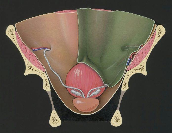
Uterine fibroids are noncancerous growths that develop in or around the uterus. When these growths form on the outer surface of the uterus, they are referred to as subserosal fibroids. Detecting such fibroids requires specific diagnostic approaches, as their location can make them less obvious during routine evaluations. A specialized health clinic employs various tools and techniques to identify those located outside the uterine wall. This article explores the structured process used by medical professionals to diagnose subserosal fibroids.
Initial Patient Consultation and Medical History Review
The diagnostic process for uterine fibroids outside the uterus begins with a comprehensive patient consultation. During this stage, healthcare providers gather detailed information about symptoms, family history, and previous medical conditions. Common indicators of subserosal fibroids include pressure or pain in the lower abdomen, frequent urination, or digestive discomfort. Patients may not always experience noticeable symptoms, making background health data especially valuable. By understanding the individual’s unique health profile, clinicians can more accurately assess the likelihood of fibroid development and determine which diagnostic steps are most suitable.
Physical and Pelvic Examination Techniques
Following the initial discussion, a physical evaluation is conducted to detect any abnormalities in the pelvic region. A pelvic exam allows the physician to feel for irregularities in the shape or size of the uterus. In some cases, large subserosal fibroids can be identified through manual palpation.
While this method provides preliminary insights, it does not offer detailed visualization of the fibroid’s exact location or size. Therefore, it serves as an early step that guides further diagnostic planning rather than a conclusive assessment. The physician may also assess for tenderness or discomfort during the exam, which can indicate the presence of fibroids pressing on surrounding structures.
Transvaginal and Abdominal Ultrasound Imaging
Imaging tests play a central role in confirming the presence of uterine fibroids outside the uterus. Transvaginal ultrasound involves inserting a wand-like device into the vagina to capture images from within, which offers a close view of the uterine structure. Alternatively, abdominal ultrasound uses a transducer moved across the lower abdomen to generate external images. Both methods utilize sound waves to visualize internal organs without exposing them to radiation. These imaging techniques help distinguish between different types and provide essential details such as size, number, and position of the uterus.
Magnetic Resonance Imaging (MRI) for Precise Localization
When more detailed imaging is required, magnetic resonance imaging (MRI) may be recommended. MRI uses powerful magnets and radio waves to produce high-resolution cross-sectional images of the pelvis. It is particularly useful in identifying subserosal fibroids and differentiating them from other pelvic masses. The clarity of MRI scans supports accurate diagnosis and helps in treatment planning. This technique can reveal fibroid characteristics that influence management decisions, such as blood supply and proximity to surrounding tissues.
Use of Hysteroscopy for Uterine Cavity Evaluation
Hysteroscopy is the insertion of a thin, lighted tube through a patient’s cervix to examine the inside of her uterus. Although primarily used to evaluate intrauterine fibroids, this procedure helps rule out other causes of symptoms when findings are inconclusive. It is typically performed in an outpatient setting and offers immediate visual feedback. While hysteroscopy cannot directly visualize subserosal fibroids, it complements other diagnostic methods by ensuring a complete assessment of all uterine structures. The procedure can also help identify any associated abnormalities, such as polyps or adhesions that may contribute to a patient’s symptoms.
Diagnostic Laparoscopy for External Fibroid Identification
Laparoscopy is a minimally invasive surgical procedure that allows direct visualization of the outer surface of the uterus and surrounding areas. A small incision is made near the navel, through which a laparoscope, a slender instrument with a camera, is inserted. This technique enables physicians to observe fibroids located externally and assess their impact on adjacent organs. Laparoscopy can also serve both diagnostic and therapeutic purposes, depending on the situation. Due to its accuracy, it remains a reliable option when other imaging methods fail to provide sufficient detail.
Analysis and Interpretation of Imaging Results
Once imaging procedures are completed, specialists review the collected data to confirm the diagnosis. Radiologists and gynecologists collaborate to interpret ultrasound, MRI, or laparoscopic images accurately. Findings are then correlated with the patient’s reported symptoms and physical examination results. This multidisciplinary approach ensures consistency and reduces the chance of misdiagnosis. A clear diagnostic summary is provided to the patient, outlining the nature of the fibroid and available options for care. The integration of clinical findings with imaging data helps modify a treatment plan that aligns with the patient’s health status and personal preferences.
A health clinic utilizes a structured and comprehensive approach to diagnose uterine fibroids outside the uterus. Each diagnostic step is designed to gather precise information about the size, location, and impact of subserosal fibroids. From initial consultations to advanced imaging techniques, medical professionals rely on a combination of tools to ensure accurate identification. These methods support informed decision-making regarding treatment options. Hence, effective diagnosis is essential for developing a personalized care plan that addresses each patient’s unique condition.


